Dr Denning welcomed the participants. He described the size of the problem of invasive aspergillosis (IA) in terms of its incidence in different patient populations:
| Lung transplantation | 17-26 % |
| Allogeneic bone marrow transplantation | 5-15 % |
| Acute leukaemia | 5-24 % |
| Heart transplantation | 2-13 % |
| Pancreas transplantation | 1-4 % |
| Renal transplantation | 0.5-10 % |
| AIDS | 0-12 % |
| Multiple myeloma (stage III) | ~4 % |
| Severe combined immunodeficiency | ~4 % |
| Solid tumour and lymphoma | ~1-3 % |
| Autologous BMT (with growth factors) | ~1 % |
| Connective tissue diseases (e.g. SLE) | ~1 % |
| Non-immunocompromised patients | <1 % |
He noted the high mortality of invasive aspergillosis (50 to100 %) even with therapy and discussed the report from Frankfurt (Groll et al., J Infection 1996; 33: 23-32) which showed a 14-fold rise (over 12 years) in the incidence of invasive aspergillosis at autopsy. He presented antifungal sales figures from 1997 from Europe, USA and Japan of which $370 M from a total of $1230 M was spent on the only two licensed drugs with activity against Aspergillus - amphotericin B and itraconazole. He emphasised that invasive aspergillosis is now the leading cause of early death in many transplant centres and has a major impact on the management of leukaemia.
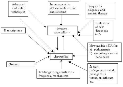
Dr Denning then presented an overview of strategies to address this problem (Figure I.). Subsequent contributions to the workshop were specifically targeted to one or more of these issues.
Finally, he informed the workshop of progress towards getting the genome of Aspergillus fumigatus sequenced. A pilot project to create and map a bacterial artificial library (BAC) of a pathogenic isolate of A. fumigatus is under consideration by Beowulf, the pathogen sequencing arm of the Wellcome Trust. This project is a collaboration initially between the Sanger centre in Cambridge and the University of Manchester. The project proposes to sequence ten clones - about 1 Mb of the estimated 30+ Mb genome. The region to be sequenced corresponds to a well characterised area of A. nidulans chromosome VIII (containing the nitrate assimilation gene cluster; crnA, niiA and niaD). The workshop unanimously agreed that a genome sequencing project was important to improve the identification of novel diagnostic antigens/targets, the development of new antifungal targets, the understanding of antifungal resistance and the understanding of the biology and pathogenicity of A.fumigatus. The sequence data could also generate new commercial enzyme or metabolic products because of the remarkable thermotolerance of A. fumigatus.
IA in neutropenia and bone marrow transplant recipients risk
factors and outcome
Dr Martino gave an overview of the size of the problem of
filamentous fungal infection in leukaemia and bone marrow
transplantation. He noted that in the group he represented (GIMEMA
group in Italy - 60 centres), 48 % of these infections occurred at
the onset of leukaemia, for example during the first course of
induction remission therapy. These infections were often fatal;
either directly because antifungal treatment failed or because
chemotherapy for the leukaemia was delayed, compromising its
treatment. Filamentous fungal infections therefore present a
serious problem in the management of leukaemia today. Dr Martino
also discussed the control of Aspergillus in the environment
and focussed on the recent descriptions of Aspergillus in
water supplies and showerheads, the significance of which is
currently unclear.
IA in solid organ transplant recipients risk factors &
outcome
Dr Pahissa presented an overview of the problems of invasive
aspergillosis in solid organ transplant recipients. He described
the group set up in Spain to study infection in these patients
(GESITRA - SEIMC). This group, comprising transplant surgeons,
transplant physicians and infectious disease physicians
collectively cares for about 1400 renal, 1700 liver, 900 heart and
60 lung transplant recipients annually. He noted that in their
experience the mortality of invasive aspergillosis varied from 27
to 77 %. He also observed that the median time to diagnosis was six
days, and that 78 % of diagnoses were made antemortem. In their
multivariate analysis the following were all independent risk
factors for death: renal insufficiency, the need for additional
surgery because of surgical complications during the
transplantation procedure, a lack of cotrimoxazole prophylaxis,
chronic rejection and retransplantation. Only the use of AmBisome
was an independent predictor of survival.
Clinical and radiological diagnosis in non neutropenic
patients
Dr Grossi described diagnostic approaches in solid organ transplant
patients with a particular emphasis on lung transplant patients.
These approaches were based on the experiences of the 12 centres in
the Italian Study Group for Fungal Infections in Thoracic Organ
Transplantation. He noted that most cases occurred two to three
months after transplantation. In lung transplant recipients their
experience of bronchoalveolar lavage for diagnosing invasive
aspergillosis is shown below:
| IA | no IA | |
|---|---|---|
| BAL positive culture (Aspergillus) | 8 | 41 |
| BAL negative culture | 0 | 398 |
Sensitivity = 100 %, specificity = 92 %
Positive predictive value = 16 %
Negative predictive value = 100 %
Some patients had multiple positive cultures and did not develop IA although almost all were given oral itraconazole. He observed that among the lung transplant recipients, 35 % of those with IA had dissemination to other organs and overall, the mortality in their hands was about 35 %.
Serological diagnosis
Dr Latgé described approaches
to serological diagnosis that have been used. He mentioned interest
in mannitol and certainly this metabolite of Aspergillus has
been detected in patients and in experimental infections. However
mannitol has not been systematically used in the clinical setting.
Antibody detection is of great use in the diagnosis of aspergilloma
and allergic bronchopulmonary aspergillosis, but has been
disappointingly negative in patients with invasive aspergillosis.
All controls have low levels of detectable antibody and so the
standard cut-off for a positive test is a one in ten dilution.
Study of lower levels of positivity or a fall off in levels as
antibody is 'consumed' were presented as possible ways forward.
Another suggestion was to improve the actual antigen(s) and
standardise these with recombinant technology.
Dr Latgé also described attempts to localise infection using directly injected optical brighteners. This needs more work but is conceptually attractive. Similarly the concept of monoclonal antibody imaging has not been developed, but is attractive.
The only marketed antigen test in Europe is Sanofi Pasteur's
Platelia sandwich ELISA. Dr Latgé remarked on its utility,
noting that the galactomannan that is detected is produced by the
fungus during infection irrespective of the host response. However
it is chemically proven that many foods, especially noodles,
contain galactomannan and this could account for the relative
frequency of false positive tests. Little or no data exist on the
usefulness of the test in non-haematology patients. He contrasted
this antigen test with the so-called G-test that is marketed in
Japan and detects 1-3, ß-D-glucan. The glucan test is likely
to be positive (it is very sensitive) in patients with some degree
of immune response as the release of 1-3, ß-D-glucan only
occurs when the fungal cell wall is damaged. Comparative tests
between ß-1-3 glucan and galactomannan have been started.
Polymerase Chain Reaction detection of Aspergillus DNA
in samples
Dr Bretagne described the PCR tests that have been developed in his
laboratory and their relative merits and problems. He outlined the
various contamination problems that can beset PCR. These include
extraneous contamination of the original sample or of the PCR mix
with Aspergillus conidia and more importantly, contamination
of the PCR mix with amplicons from prior reactions. Several
stringent quality control measures were advocated to prevent false
positive results, including the systematic use of
uracil-DNA-glycosylase (UNG) which cleaves any contaminating
amplicons prior to amplification. Dr Bretagne also advised against
the use of nested PCR which is more liable to false positives. He
also noted that inhibitors of the amplification reaction could
yield false negative results. This inhibition can be detected by
amplifying an internal control reaction in each sample tested.
Finally Dr Bretagne argued that only high quality samples should be
analysed to avoid interpretation problems.
PCR and T cells responses to Aspergillus
Dr Hebart described his group's results with a PCR test that
firstly amplifies DNA from all pathogenic fungi and then utilises a
hybridisation step to differentiate the underlying fungal pathogen.
He presented the results of a prospective screening program on
blood samples from recipients of an allogeneic stem cell
transplant. In patients with proven and probable invasive
aspergillosis, PCR positivity preceded clinical signs of IA by a
mean of 21 days. PCR positivity in the later post transplant period
was found to be associated with a graft versus host disease (GvHD)
and corticosteroid treatment, major risk factors for IA. Based on
these results, the group led by Drs Hebart and Einsele has started
a multicenter trial comparing PCR-based versus empirical antifungal
therapy after allogeneic stem cell transplantation.
Dr Hebart also described preliminary results on Aspergillus-specific lymphoproliferation in healthy volunteers and patients with IA. Lymphoproliferative T-cell responses were highest in patients surviving IA, whereas these responses were not detectable in recipients of an allogeneic stem cell transplant in the early post-transplant phase (up to day + 100). Further details are warranted.
How does immunosuppression, radio- and chemotherapy affect
epithelium and immune cells?
Dr Poynton noted that little research
had addressed the issue of local and systemic damage wrought by
chemotherapy and radiotherapy relative to a) the number of patients
treated and b) the size of the problem. In the context of acute
induction therapy for leukaemia, he described experiments using
mannitol and lactulose to quantify damage to the gut. Mannitol is
actively reabsorbed in the normal gut, but lactulose absorption is
a measure of damage to intercellular boundaries. A ratio of the two
allows direct quantification of gut damage, which is considerable
in the few patients they have studied so far. Further work has
shown that the level of endotoxin in blood is directly related to
gut damage. Thus fever in neutropenia could reflect gut damage and
risk of infection as much as infection itself. Glutamine is
somewhat protective of epithelial damage and could be important in
protecting patients from aspergillosis or may be a marker of
damaged epithelial surfaces.
Dr Poynton went on to discuss the relationship between cytokine
expression and cytokine levels using a small dataset he had
collected. It was already apparent that there was no relationship
in the cells they examined (IL4 &IL12 positive CD4+ cells).
Finally, he discussed the possible role of nitrous oxide (NO) and
speculated that the anergy noted just before death could be
attributable to 'disregulation of NO'.
How do phagocytes kill Aspergillus, an extracellular
pathogen?
Dr Roilides gave an overview of the killing of Aspergillus
conidia and hyphae by the oxidative and non-oxidative mechanisms of
phagocytes (neturophils, monocytes, macrophages). He noted that the
subcellular and biochemical mechanisms of phagocytes still remain
unknown. He also emphasised the enhancing effects of hemopoietic
and Th1 cytokines on certain antifungal activities of phagocytes
against Aspergillus. He went on to discuss the unknown
importance of non-oxidative enzymes and other substances including
NO and its possible role in the killing of conidia by macrophages.
Antimicrobial peptides of leukocytes (e.g. defensins, lactoferrin
lysozyme, bactericidal permeability-inducing protein, calprotectin,
protegrins, cecropins, etc) have not been investigated to any
degree, but are probably important. Xoma, a California-based
company, has developed a series of such compounds one of which has
substantial in vivo activity in an animal model of
aspergillosis. In other fungi substantial work has been done on the
interactions of phagocytes, hemopoietic cytokines (e.g. granulocyte
colony stimulating factor) and antifungal drugs (e.g. azoles), a
synergism which may be very important in patients with fungal
infections. However, similar work has not been performed with
Aspergillus or other antifungal drugs (e.g. lipid formulations
of amphotericin B, echinocandins, pneumocandins, etc).
Cytokine responses to Aspergillus infection
Dr Romani spoke about the substantial variation in susceptibility
to invasive aspergillosis in different strains of mice. In a murine
model of invasive aspergillosis, production of Th1 and Th2
cytokines were different in mice resistant or susceptible to the
infection. Development of protective acquired immunity required
IL-12 and was associated with the production of IFN-g from Th1 cells upon
in vitro antigen-specific stimulation. In contrast,
production of IL-4 and IL-10 by antigen-activated Th2 cells was
associated with disease progression and down-regulation of
protective Th1 immunity. IL-4 neutralisation or IFN-g administration
had a curative effect in invasive aspergillosis, indicating the
critical role of these cytokines in the pathogenesis of this
infection. Similar to observations in mice with invasive
aspergillosis, innate immunity and the generation of Th1 cytokines
play an essential role in host defenses against invasive pulmonary
aspergillosis (IPA). Resistance to infection was associated with
the unimpaired innate antifungal activity of pulmonary phagocytic
cells. This was concomitant with high-level production of
TNF-a and IL-12 and the
presence of interstitial lymphocytes producing IFN-g. Conversely, production of TNF-a and IL-12 was down-regulated in highly
susceptible mice showing defective innate antifungal immunity and
high-level production of IL-4 and IL-10 by lung lymphocytes.
Resistance was increased in susceptible mice upon local IL-4 or
IL-10 neutralisation or IL-12 administration
Circumstantial evidence in humans indicates that dysregulated production of Th cytokines may contribute to the pathogenesis of IPA. These include the pathogenetic role IL-10 may have in aspergillosis in patients with HIV infection, the occurrence of pulmonary aspergillosis in patients with the hyperimmunoglobulin E syndrome and the recently reported ability of human lymphocytes to respond to A.fumigatus conidia in vitro with activation, proliferation, and production of Th1 cytokines. Should this last finding be extended to patients, it may be possible to identify persons at increased risk of aspergillosis by the quality of the Th cell response to in vitro Aspergillus antigens.
Genetics of cytokine responses to micro-organisms
Dr Ollier gave a broad introduction to genetic factors determining
immune response. He spoke of polymorphisms in the promoter regions
of TNF-a and the correlation
between IL-10 polymorphisms and circulating levels of this
cytokine. He summarised the data on mannose binding lectin (MBL)
which binds to Aspergillus, is important in the clearance of
pathogens and immune complexes and is an activator of the alternate
complement cascade. Five mutations have been described, two in the
promoter region (H/L and Y/X) and three in the open reading frame
(at codons 52, 54 and 57). These lead to more or less functional
circulating MBL. The codon 54 mutation is present in 16 % of
caucasians and is homozygous in 2 %. Combinations of promoter
mutations lead to higher or lower serum MBL levels so that the HY
genotype has high levels and the LX genotype is essentially
deficient. MBL deficiency leads to an increased frequency of
bacterial infections particularly otitis media and pneumonia. No
one has investigated its possible role in any of the forms of
aspergillosis.
NIH funding
Dr Dennis Dixon, Chief of the
Bacteriology and Mycology Branch of the NIH/NIAID/DMID, outlined
the basic approaches to NIH funding and the internal structures of
the NIH. He indicated that funding for fungal projects could come
from several sources within the NIH, although most is handled, at
least in part, by the Division of Microbiology and Infectious
Diseases. Over the last three years (1995-1997) funding for the
mycology program within that Division had risen to around $15 M
annually. The program goals for mycology were:
1) Diagnosis, treatment and prevention of serious mycoses
2) Mechanisms of pathogenesis
3) Improvements in treatment through basic and clinical studies,
including comparative clinical trials.
He identified the gap areas in mycology as:
1) Training in contemporary mycology
2) Research on fungal vaccines
3) Research on Aspergillus and other moulds
4) Fungal genome research on human pathogens
He noted that the NIH would be very supportive of any EU effort
to sequence the A.fumigatus genome. The NIH website carries much information -
including reports on mycology
workshops
In vitro and animal models for the study of
pathogenesis
Dr Latgé described the animal models that have been used to
study virulence. He summarised the findings to date as essentially
negative, despite the considerable investment of time and resources
in generating and evaluating isogenic single and double mutants.
Many questions remain: for example, the fate of conidia, the
different steps in the infection process and why there are so many
different manifestations of aspergillosis.
Dr Latgé also summarised the issues regarding adhesion. Conidia adhere to several separate components of the external milieu including collagen, complement, fibrinogen, laminin, fibronectin, surfactant, and a phagocyte receptor. The conidial cell wall has three identified binding components, rodlet proteins, pigment and lectin-like coating. Binding to fibrinogen and laminin is for example, sialic acid dependant. Given this multiplicity of possible binding combinations, it may be unrealistic to consider blocking it as a therapeutic avenue.
Dr Latgé then addressed the question of the mechanisms of conidial killing by alveolar macrophages and concluded that we do not know how they kill conidia. The same is almost true with neutrophils.
He raised a critical point for future work, namely a definition of virulence. Most workers have been using increased or reduced mortality in a mouse model as the definition, but this is a crude approach and subject to much experimental error. He recommended that such a definition be agreed in the near future. Identification of genes differentially expressed during infection would avoid any wrong a priori selection of putative virulence factors. A program to approach this strategy is presently being undertaken at the Pasteur Institute.
Single gene disruption in Aspergillus
Dr Monod presented the different strategies available to obtain
single disruption events in A. fumigatus. Two drug
resistance markers (hygromycin and phleomycin) have been used to
successfully transform A. fumigatus. Dr. Monod gave a list
of the different genes which have been disrupted to date including
various proteases, catalases, ribonuclease and hydrophobin. Some
experimental data supported a putative role for these proteins
during infection. Unfortunately, in a mouse model of IA, no
differences were seen between the mutants and parental strains.
These results called into question the animal model used to test
the virulence of A. fumigatus strains and raised again the
question of a polygenic nature of virulence in this opportunistic
fungus.
Dr. Monod also introduced us to Invitrogen's Pichia pastoris system used successfully for the production of recombinant proteins of A. fumigatus. Pichia pastoris respects the glycosylation of the protein and allows the production of high concentrations of protein (0.1 mg/ml) which can be secreted in the culture medium.
New molecular approaches
Dr Jeremy Brown presented an overview
of the restriction enzyme mediated integration (REMI) approach to
virulence gene identification and described his work over three
years. The signature tagged mutagenesis - restriction enzyme
mediated integration (STM-REMI) approach allows the production of a
bank of mutants by unique insertion of a resistance marker at a
random site in the A. fumigatus genome. Every mutant is
tagged at a different locus which facilitates simultaneous testing
of 96 different mutants in the same mouse. Any mutant(s) which have
not grown in the mouse must have a gene essential for virulence
disrupted and these genes can be cloned owing to their unique
flanking sequence tag. He described how 4648 mutants were tested
(56 pools of mutants on three to five mice per pool). From this
large screen, two mutants of interest were found. The first was
less virulent and had a disruption event in a small ORF; this ORF
revealed no homologues. The second mutant was a para-amino benzoic
acid (paba) auxotroph and was fully avirulent (not germinating in
the mouse lung). These results also questioned the importance of
single mutations in the study of A. fumigatus virulence and
would suggest that virulence is multifactorial.
Identifying new candidates as virulence determinants
biochemically
Dr Ruchel presented what little was known about secreted enzymes in
Aspergillus. Various secreted or cellular proteins of the
opportunistic pathogen A.fumigatus have been suspected of
being pathogenicity factors. However, targeted deletion of
individual corresponding genes has met with discouraging results
from subsequent experimental infections. The normal habitat of
A.fumigatus is not the warm blooded host, but rather moist
organic matter in the environment. Therefore, the existence of such
"virulence factors", expressed by the fungus for survival in the
human host, is questionable. However, we assume that fungal
proteins may still warrant attention because proteins critical in
essential biochemical cellular pathways or for fungal growth in the
host could be targets for future pharmacotherapy. These proteins
should be enzymes, and thus targets of inhibitors and must differ
from human proteins. Candidates are enzymes of the cell wall, of
the vacuole, or of other cellular compartments which are specific
to fungi. Dr Ruchel favoured vacuole-associated enzymes.
Mycotoxins and Aspergillus fumigatus
Dr Galtier described what was known about the toxins of
Aspergillus. He noted that the tryptostatins (A, B) inhibit
cell cycle progression at M phase, but the relative toxicity of
each is not known. He noted that there were four tryptoquivalines
all with low toxicity potential. In contrast, verruculogen was very
toxic causing tremors and fits. Its mechanism of action is not
fully characterised, but it may interact with the neurotransmitter
- amino butyric acid (GABA). Similar but in decreasing order of
toxicity are the fumitremorgens A, B, C and TR-2 and C.
Dr Galtier then focussed on gliotoxin which is produced by A.fumigatus, A.flavus and C.albicans. A six day culture of each organism in Eagle's or YES medium with 5 % fetal calf serum will yield relatively large quantities of gliotoxin. It has been found in necrotic bovine udders, peritoneal washings in infected mice and in lung tissue of turkeys in very variable concentrations. It is rapidly taken up by cells and causes reduced phagocytosis and apotosis probably via multiple mechanisms including RNA polymerase inhibition, inactivation of intracellular proteins and possibly the generation of intracellular hydrogen peroxide. Gliotoxin may also cause damage to epithelial cells. Dr Galtier outlined the probable synthetic pathway for gliotoxin and noted that the pertinent enzymes were not known or cloned.
Epidemiology of resistance in Aspergillus
Dr Rodriguez-Tudela gave a short summary of progress in antifungal
susceptibility testing over the last decade. He outlined the
impressive progress towards standardisation in yeast susceptibility
testing by the NCCLS subcommittee in the US. He outlined problems
with the methodology and breakpoint interpretation including the
testing of amphotericin B and the poor growth of some yeasts
(especially Cryptococcus) in RPMI without glucose
supplementation. He then spoke about 'problem species' and the need
to standardise methods and conduct contemporary surveys of
resistance in Aspergillus. He mentioned the formation of a
new European Committee for Susceptiblity Testing (EUCAST)
subcommittee on antifungal susceptibility testing that would
address many of these issues.
Molecular basis for resistance in Aspergillus
Dr Denning described what little was known about itraconazole
resistance in Aspergillus fumigatus. Two mechanisms of
resistance were postulated a) overexpression of an efflux pump
leading to reduced intracellular concentrations of itraconazole and
b) mutation of the itraconazole target, 14a -demethylase. Work proceeds on cloning the
relevant genes. Dr Denning then discussed the issue of cross
resistance as animal models and in vitro studies have
indicated a degree of cross resistance with SCH 56592 (an azole in
phase II development), but no cross resistance to voriconazole (an
azole in phase III development). The structure SCH 56592 is similar
to that of itraconazole, whereas that of voriconazole is not.
He then showed some animal model data consistent with amphotericin B resistance in one isolate of Aspergillus. He mentioned a recent paper by Dr Lass Florl in the Journal of Antimicrobial Chemotherapy which purported to be able to separate, with a breakpoint of >2 mg/ml, amphotericin B resistant isolates from susceptible ones. Those patients infected with the 'resistant' isolates, all died. There was some missing data from the paper, but overall it seems possible that amphotericin B susceptibility testing will soon be possible for Aspergillus. The mechanism of action of amphotericin B in Aspergillus is unknown.
Dr Denning then bid farewell to all the participants and thanked them for their contributions and attendance.
Prepared by D.W. Denning and J.P. Latge, March 1999
Participants
| Dr David W Denning
(Go to the link for up-to-date details) Head, Section Infection Diseases Department of Medicine, University of Manchester North Manchester General Hospital Delaunays Road Crumpsall Manchester, M8 5RL |
Tel: +44 161 720 2734 Fax: +44 161 720 2732 e-mail: Go here for up-to-date contact details |
| Dr Jean-Paul Latge
Head, Laboratoire des Aspergillus Unite de Micologie Institute Pasteur 25 rue due Docteur Roue 75724 Paris Ceden 15 France |
Tel: 33 145 688 225
Fax: 33 145 613 3419 e-mail: jplatge@pasteur.fr |
| Professor Pietro Martino
Cantro Transfusionale Universita "La Sapienza" Via Chieti 7 00100 Roma Italy |
Tel: 39 06 85795515
Fax: 39 06 44241984 e-mail: martino@bce.med.uniromal.it |
| Dennis Dixon
Chief, Bacteriology & Mycology Branch Division of Microbiology & Infectious Diseases NIH/NIAID Bethesda, MD 20892 USA |
Tel: 001 301 496 7728
Fax: 001 301 402 2508 e-mail: dd24@nih.gov |
| Dr Albert Pahissa
Consultant Infectious Diseases Hospital General Vall d'Hebron Barcelona Spain |
Tel: 34 93 274 6057
Fax: 34 93 274 6057 e-mail: pahissa@vhebron.es |
| Dr Chris Poynton
Department of Haematology University of Wales College of Medicine Health Park Cardiff CF4 4XN |
Tel: +44 01222 747747
Fax: +44 01222 744655 e-mail: poynton@cardiff.ac.uk |
| Professor Paulo Grossi
Division of Infectious Diseases Istituto di Clinica delle Malattie Infettive IRCCS San Matteo University of Pavia Via Taramelli 5 27100 Pavia Italy |
Tel: 39 382 525308
Fax: 39 382 423320 e-mail: pgrossi@matteo.pv.it |
| Dr Emmanuel Roilides
Assistant Professor Department of Paediatrics Aristotle University Thessaloniki Greece |
Tel: 30 31 892 447
Fax: 30 31 852 925 e-mail: roilides@med.auth.gr |
| Dr Stephan Bretagne
Consultant Laboratoire de Parasitologie-Mycologie Hopital Henri Mondor Creteil France |
Tel: 33 1 49 80 3641
Fax: 33 1 49 81 3601 e-mail: bretagne@univ-paris.12.fr |
| Dr Reinhard Ruchel
Hygiene Institut der Georg-August-Universitat Gottingen Gottingen Germany |
Tel: 49 551 395 855/7
Fax: 49 551 395 860 |
| Dr Juan-Luis
Rodriguez-Tudela
Unidad de Microbiologia Centro Nacional de Microbiologia Instituto de Salud Carlos III Ctra. Majadahonda-Pozuela km2 28220 Spain |
Tel: 34 91 5097961
Fax: 34 91 5097966 e-mail juanl.rodriguez-tudela@isiii.es |
| Dr Jeremy Brown
Department of Thoracic Medicine University College London Hospitals Middlesex Hospital Mortimer Street London W1N 8AA |
Tel: +44 171 380 9005
Fax: +44 171 637 5809 |
| Dr Michel Monod
Head Laboratoire de Mycologie Department of Dermatology Cantonal Hopital Universitaire de Vaud Lausanne Switzerland |
Tel: 41 21 314 0376
Fax: 41 21 314 0378 |
| Dr Luigina Romani
Assistant Professor Department of Experimental Medicine & Biochemical Science University of Perugia School of Medicine Perugia Italy |
Tel: 39 75 585 3411
Fax: 39 75 585 3400 e-mail lromani@egeo.unipg.it |
| Professor William Ollier
Professor of Molecular Immunogenetics Faculty of Medicine, Nursing & Dentistry University of Manchester Oxford Road Manchester M13 9PT |
Tel: 44 161 275 5622
Fax: 44 161 275 5043 e-mail: bill@fs1.ser.man.ac.uk |
| Dr Ingrid Wünning
Senior Scientific Secretary for Biomedical Sciences European Science Foundation 1 Qual Lezay-Marnésia 67080 Strasbourg Cedex France |
Tel: 33 388 7671 18
Fax: 33 388 3705 32 e-mail: emrc@esf.org |
| Dr Holger Hebart
Med Klinik II Department of Haematology & Oncology Eberhard-Karls-Universitat Tubingen 72076 Tubingen Germany |
Tel: 49 7071 2983726
Fax: 49 7071 293179 e-mail: hrhebart@med.uni-tuebingen.de |
| Dr Pierre Galtier
Laboratoire de Pharmacologie-Toxicologie INRA BP3, 180 Chemin de Tournefeuille F 31931 Toulouse France |
Tel: 33 561 285146
Fax: 33 561 285310 e-mail: pgaltier@isard.toulouse.inra.fr |
| Dr Jose Antonio Gutierrez
Fuentes
Director General del Instituto de Salud Carlos III Sinesio Delgado 6, pabell=F3n 3 28029 Madrid, Spain |
Fax: 34 91 3877832 |
| Dr Alberto Corona
Division of Infectious Diseases Istituto di Clinica delle Malattie Infettive IRCCS San Matteo University of Pavia Via Taramelli 5 27100 Pavia Italy |
Tel: 39 382 525308 Fax: 39 382 423320 |
| Invited participants unable to attend
Professor Ben de Pauw Department of Haematology University Hospital PO Box 9101 Niejmegen NL 6500 HB The Netherlands |
Tel: 31 243 542 808 Fax: 31 243 542 080 e-mail B.dePauw@HEMA.AZ.NL |
| Professor Steven Kelly
Professor of Biological Sciences University of Aberstwyth Wales SY23 3DA |
Tel: 44 1970 621515
Fax: 44 1970 622 622350 e-mail: les@aber.ac.uk |
| Professor Peter Cole
Professor of Thoracic Medicine Royal Brompton National Heart & Lung Hospital University of London London SW3 6LR |
Tel: 44 171 351 8326
Fax: 44 171 351 8338 e-mail: a.burling@ic.ac.uk |
| Professor Hermann Einsele
Professor of Haematology Department of Haematology & Oncology Eberhard-Karls-Universitat Tubingen 2076 Tubingen Germany |
Tel: 49 7071 298 2726
Fax: 49 7071 293671 e-mail: hneinsel@uni-tuebingen.de |
| Dr Paul Verweij
Consultant Department of Microbiology University Hospital Nijmegen The Netherlands |
Tel: 31 14 361 4356
Fax: 31 24 354 0216 e-mail: p.verweij@mmb.az.nl |
| Professor Michel Glauser
Professor of Infectious Diseases University Hospital Lausanne 1011 Switzerland |
Tel: 00 41 21 314 1010
Fax: 00 41 21314 1018 e-mail: Sylviane.Bovey@chuv.hospvd.ch |
| Dr Guy Tronchin
Laboratoire de Parasitologie Mycologie Centre Hospitalier Universitaire Angers France |
Tel: 33 0241 35 34 72
Fax: 33 0241 35 36 16 |
| Dr Per Ljungman
Associate Professor of Medicine Huddinge University Hospital Kurolinska Instituter S.14186 Huddinge Sweden |
Tel: 468 746 1000
Fax: 468 774 8725 e-mail: per.ljungman@medhs.ki.se |
| Professor E R Moxon
Professor of Paediatric Infectious Diseases Institute of Molecular Medicine University of Oxford John Radcliffe Hospital Headington Oxford OX3 9DU |
Tel: 01865 221074
Fax: 01805 220479 e-mail: richard.moxon@paediatrics.ox.ac.uk |